Cell structure
Cell Structure
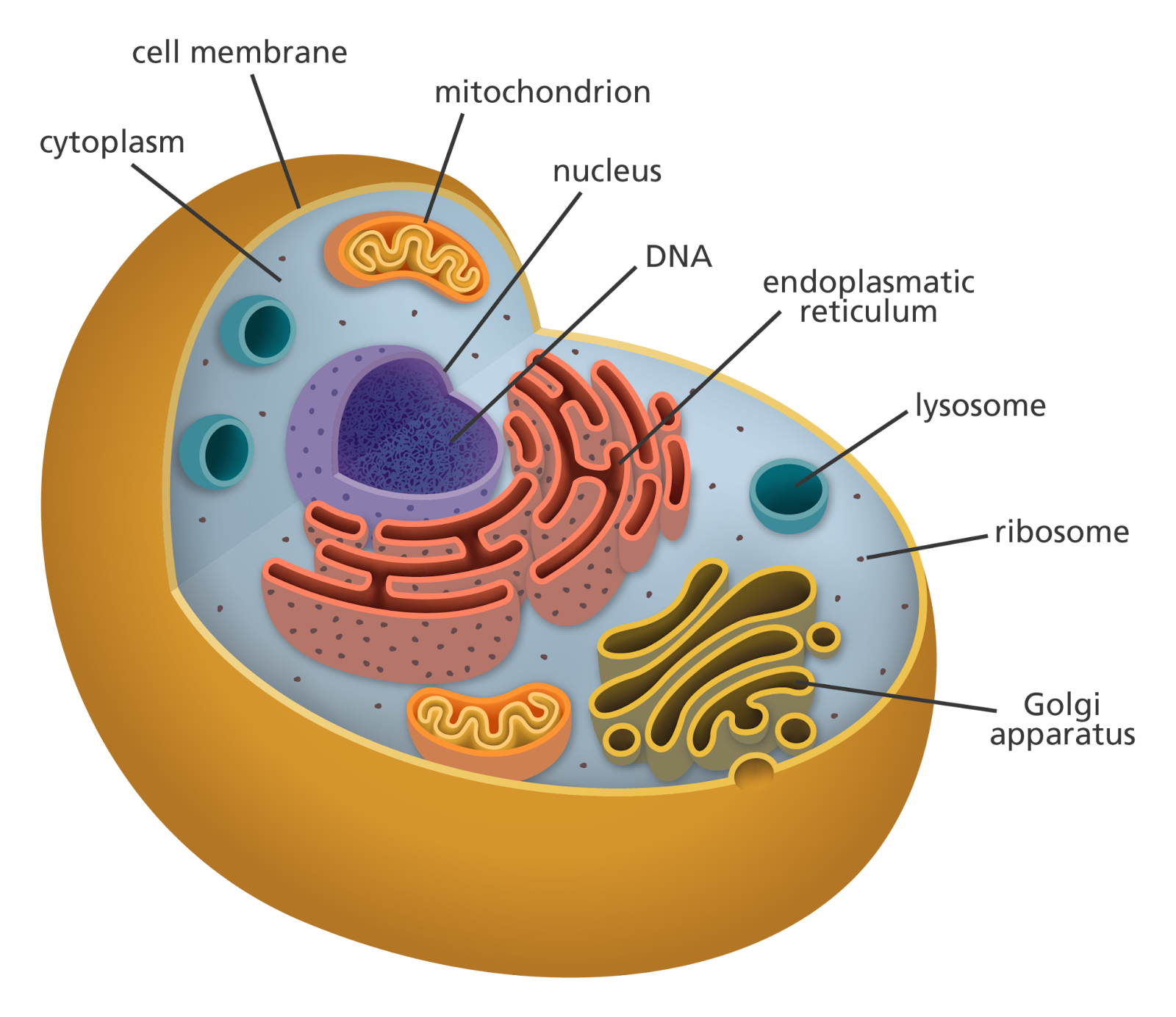
Take a good look at your hand. It looks solid and continuous. But if we could zoom in millions of times, you would see that your hand is made of tiny living units called cells. Every plant, animal, fungus, and bacterium is built from cells.
The cells of all living things are, in a sense, like the atoms of all matter. Cells are the basic structural and functional units of all living organisms. Once we know the structure of a cell, it is easier to understand the life processes that all living things share.
The Discovery of Cells
The history of cell study began in 1665 when Robert Hooke used a microscope to look at cork. He described the cells (boxlike structures) that he saw. Later, other scientists such as Matthias Schleiden and Theodor Schwann proposed the Cell Theory.
Cell Theory
Cell Theory states:
- All living organisms are made of one or more cells.
- The cell is the smallest unit of structure and function.
- All cells come from other cells.
This is one of the many theories of modern biology.
Prokaryotic and Eukaryotic Cells
There are two main types of cells: prokaryotic and eukaryotic.
Prokaryotic Cells
Prokaryotes are simple and smaller. They have no nucleus and their genetic materials are in the cytoplasm. An example of a prokaryotic cell is Escherichia coli.
Main features:
- No nucleus
- Circular DNA
- No organelles
- Ribosomes are smaller and are called 70S
- Peptidoglycan in the cell wall
Prokaryotes can have other features like:
- Flagella for movement
- Pili for attachment
- Plasmids (small circular DNA)
Eukaryotic Cells
Eukaryotic cells are the opposite. They are larger and have a more complex structure. They have a true and Membrane-bound nucleus. Plants, animals, fungi, and protists are all eukaryotes.
Main features:
- They have a membrane-bound nucleus
- DNA that is linear and is associated with proteins
- They have membrane-bound organelles
- Their Ribosomes are called 80S and are larger
- Compartmentalization of functions
Later on, we will discuss the differences that exist between animal and plant cells.
Cell Size
Cells, on average, are 1–100 micrometers (µm) in size. Their size is determined by the amount of surface area they have in relation to their volume.
Cells also have to exchange materials, which is more difficult and less efficient the larger a cell is. Cells can only grow to a certain limit, which is why they remain small. Once they have reached their limit, they have to divide to produce more cells.
Microscopy
Microscopy is the study of objects that are too small to be seen by the naked eye. Cells are studied using various types of microscopes.
Light Microscope
Light microscopes provide a magnification of about 1000–1500 times by utilising light. This type of microscope is useful when studying living cells and tissues.
Electron Microscope
Rather than light, electron microscopes use streams of electrons to generate images. This means that they have much higher resolution than light microscopes.
There are two major types of electron microscopes:
- TEM (Transmission Electron Microscope) - demonstrates internal structures.
- SEM (Scanning Electron Microscope) - demonstrates surface structures.
The details of certain organelles are only seen by means of electron microscopes.
Cell Membrane Structure
The membrane of a cell in different types of cells is described by the Fluid Mosaic Model introduced by Singer and Nicolson.
The membrane consists of:
- A phospholipid bilayer
- Proteins
- Cholesterol (only in animal cells)
- Glycoproteins and glycolipids
Phospholipids are made of:
- A hydrophilic (water-loving) head
- A hydrophobic (water-fearing) tail
They are arranged in two layers with the tails of the phospholipids facing one another in the center of the membrane. This enables the membrane’s fluidity due to the ability of the phospholipids to move from side to side. It’s mosaic in appearance due to the proteins scattered throughout it.
The membrane has the following functions:
- Controls what goes in and out of the cell
- Recognition of the cell
- Communication
- Keeps the cells adhered together
Organelles & Structures
Nucleus
The nucleus is known as the control center of the cell since it holds the cell’s genetic material (DNA).
The structure contains:
- A double membrane (nuclear envelope)
- Nuclear pores
- A nucleolus (where ribosomes are made)
The nucleus has its DNA stored in the structures known as chromosomes. The DNA is the only material that can control the building of proteins and regulating of ALL activities of the cell.
Cytoplasm
The cytoplasm is the gel-like material that fills the cell. It has within it the enzymes and nutrients, as well as the organelles. It is the site for many of the cell’s metabolic reactions.
Ribosomes
Ribosomes are structures that are made of proteins and RNA and are the main site of protein synthesis. They can be classified into two types:
- Free ribosomes, which are found in the cytoplasm
- Bound ribosomes, which are attached to the rough endoplasmic reticulum (ER)
Prokaryotic ribosomes are called 70S ribosomes, while eukaryotic ribosomes are known as 80S ribosomes.
Endoplasmic Reticulum (ER)
The endoplasmic reticulum is a membranous organelle that consists of a series of folds that looks like a net. It is classified into two types:
- Smooth Endoplasmic Reticulum (S-ER) - which has no ribosomes attached to it and is responsible for the synthesis (building) and transport of lipids.
- Rough Endoplasmic Reticulum (R-ER) - which has ribosomes attached to it and is responsible for the same functions as the S-ER, but with the proteins.
Smooth ER Functions: Smooth ER is a network of tubules that does not have ribosomes. Its function is to synthesize lipids, detoxify chemicals, and store calcium ions.
Golgi Apparatus
The Golgi apparatus performs the functions of modifying, sorting, and packaging proteins from the ER. The proteins arrive in vesicles at the Golgi and are modified, then sent to the final destination.
Mitochondria
Mitochondria are the main site of aerobic respiration, which is how cells produce ATP. This is also why they are called the powerhouse of the cell.
Structure:
- Double membrane
- Inner membrane folded into cristae
- Matrix inside
Mitochondria possess their own DNA and ribosomes, which is one of the reasons the endosymbiotic theory is supported.
Endosymbiotic Theory
The endosymbiotic theory, which was developed by Lynn Margulis, posits that the mitochondria and chloroplasts of cells were originally autonomously replicating prokaryotic cells that were taken in by others.
Evidence of this theory includes:
- Double membranes
- Circular DNA
- 70S ribosomes
- Independent replication
Chloroplasts
Chloroplasts are the site of photosynthesis in plant cells.
Structure:
- Double membrane
- Stacks of thylakoids called grana
- Stroma (fluid region)
Chloroplasts also hold chlorophyll, allowing them to absorb the light energy.
Lysosomes
Lysosomes have digestive enzymes that dismantle cells’ waste, damage organelles, and attack pathogens. This organelle is more prevalent in animal cells.
Vacuoles
Vacuoles are membrane-bound sacs.
In the cells of plants and some bacteria:
- A large central vacuole
- Maintains turgor pressure
- Stores water and nutrients
In the cells of animals, the vacuoles are smaller and less numerous.
Cell Wall
Cell walls help plant cells provide support and protection. Cell walls are composed of cellulose. In some bacteria, the cell walls are also composed of cellulose, but it is called peptidoglycan. Animal cells do not have cell walls.
Cytoskeleton
The cytoskeleton is made of microtubules, microfilaments, and intermediate filaments.
The cytoskeleton has three functions:
- Maintains cell shape
- Cell movements
- Intracellular transport
Cilia and Flagella
Some cells have structures that help them move.
- Cilia are short and numerous
- Flagella are long and less numerous
In eukaryotic cells, cilia and flagella have a 9 + 2 arrangement of microtubules. In prokaryotic cells, cilia and flagella have a different arrangement.
Comparison of Plant and Animal Cells
In Plant Cells
- a cell wall
- chloroplasts
- a large central vacuole
- a fixed shape
In Animal Cells
- no cell wall
- no chloroplasts
- smaller vacuoles
- flexible shape
Both types of cells have a nucleus, mitochondria, ER, Golgi apparatus, ribosomes, and cell membranes.
Cell Specialization
In multicellular organisms, cells specialize. This means cells adapt to perform different functions.
Examples include:
- Red blood cells - distribute oxygen
- Nerve cells - send impulses
- Root hair cells - take in water
When cells separate different tasks, they become more efficient. Because of this, specialized cells are able to help more complex organisms exist.
Compartmentalization
One of the benefits of eukaryotic cells is having compartmentalization. Each organelle is able to focus on different parts of the process. This increases efficiency and stops various reactions from interfering with one another.
For example:
- Enzymes in lysosomes are digested safely inside membranes.
- Organized respiratory reactions are kept in mitochondria.
This is why compartmentalization is a huge evolutionary advantage.
Cell Structure and Function
Function and Structure are intertwined in biology. Mitochondria have folded membranes to help them produce more ATP. Nerve cells are designed to help expedite the process of signal transmission.
Essentially, the proper understanding of how cells are designed, it gives us an idea of how life exists on a small, microscopic scale.
Conclusion
Life’s building blocks are the cells. These cells, whether they are simple cells like the prokaryotic cells or more complex cells like the eukaryotic cells, are very organized and structured. Because of the discovery of cells, modern biological research, and the development of microscopy, a very complex and detailed structure inside small units has been revealed.
In Biology, cell structure provides the foundation for understanding metabolism, genetics, physiology, and evolution. Understanding the structure of cells allows us to understand the mechanisms of life in organisms.